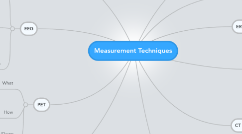
1. PET
1.1. What
1.1.1. Positron emission tomography
1.1.2. Functional processes
1.2. How
1.2.1. Labeled DFG
1.2.1.1. Used in metabolism
1.2.1.2. Shows where is used
1.3. Up/Down
1.3.1. Expensive
2. fMRI
2.1. What
2.1.1. Brain activity
2.2. How
2.2.1. Blood flow
2.2.2. Magnetic difference between oxygen rich and poor blood
2.3. Up/Down
2.3.1. Noise
2.3.1.1. Thermal
2.3.1.2. System
2.3.1.3. Physiological
2.3.1.4. Random neural activity
2.3.1.5. Metal strategy and behavior differences
3. EEG
3.1. What
3.1.1. Electrical activity
3.2. How
3.2.1. Electrodes on scalp
3.2.2. Current on scalp
3.2.3. Thought to measure extracellular activity like dendritic activity
3.3. Up/Down
3.3.1. Very poor spatial resolution
3.3.2. Very good temporal resolution
4. MEG
4.1. What
4.1.1. Magnetic activity
4.2. How
4.2.1. Sensitive magnetic detectors
4.2.1.1. SQUID - superconducting quantum interference devices
4.3. Up/Down
4.3.1. Needs magnetically shielded room
5. CT Scan
5.1. What
5.1.1. Ability to block X ray
5.1.1.1. Density
5.1.2. 3D from 2D
5.1.3. Computer tomography
5.2. How
5.2.1. X ray slices
5.2.2. Slices into model
5.3. Up/down
5.3.1. High contrast resolution
5.3.2. More accurate than radiographs
5.3.3. Can cause DNA damage and cancer
6. TMS
6.1. Magnetic de/hyperpolarisation
7. MRI
7.1. What
7.1.1. 3D image of tissue
7.2. How
7.2.1. Magnetic resonance
7.3. Up/Down
7.3.1. Good for soft tissue
7.3.2. Non ionising radiation
