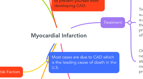
1. Most cases are due to CAD which is the leading cause of death in the U.S.
2. Signs and Symptoms
2.1. Sometimes there are no signs and symptoms but most commonly the patient might experience chest pain that radiates to the neck or arms or epigastric pain such as acid reflux pain. Dyspnea and fatigue are other symptoms
2.2. When the patient experiences an MI, the body tries to go into survival mode. You might see an increase or decrease in blood pressure and heart rate or cardiac arrythmias.
3. Treatment
3.1. Reperfusion therapy is #1 this could be taking the patient into the cath lab for PCI and stenting or fibrinolytic medications such as TPA or TNK.
3.1.1. Lifestyle Modifications will be recommended such as healthy eating and quitting smoking and drinking alcohol.
3.2. Treatment of pain with opioids such as morphine and nitrates, such as nitroglycerin depending on the area of MI. Aspirin given for protection of the heart. Treatment of anxiety and oxygen as needed.
3.2.1. After the patient is discharged, they might need a blood thinner such as plavix if a stent was placed. These patients will likely be placed on a cholesterol medication such as a statin. They will also be required to take Aspirin daily.
3.3. Other medications given as needed such as antihypertensives, atropine for low heart rate, or medications to increase the blood pressure.
3.3.1. Referrals will be made with cardiology for future treatment and resources as well as social work consults to ensure the patient can afford treatment.
4. Pathophysiology and Eitology
4.1. Caused from a decrease in blood flow to a portion of the myocardium. Most primary cause is a blood clot that breaks off and causes an occlusion.
4.1.1. An MI can trigger an inflammatory response in the heart. This inflammatory response plays a big role in how the heart will heal after a cardiac event and can lead to arrythmias and adverse cardiac remodeling.
4.2. When the myocardium becomes blocked, it lacks oxygen which cause tissue necrosis and death. Necrosis starts in the sub-endocardium, then goes to sub-epicardium.
4.2.1. Complications could include arrythmias, inability to perform surgery if unstable, possible need to do open heart surgery, or death.
5. Risk Factors
5.1. Modifiable: Smoking, Obesity, High lipid and cholesterol levels, Hypertension, Type 2 diabetes, Alcohol consumption, Decreased daily activity, Bad eating habits
5.2. Non-Modifiable: Advanced age, Male gender, genetics
6. Diagnostics
6.1. The patient needs an EKG to look at heart rhythms and ST changes, Serial troponin lab tests and a chest X-ray. Echocardiograms can be performed to look at an affected heart after an MI.
6.1.1. You might notice an increase in the patients WBC due to trauma and possible electrolyte abnormalities such as potassium and sodium.
